anatomy physiology lab manual answers
Anatomy & Physiology Lab Manual Answers: A Comprehensive Guide
Fundamentals of Anatomy & Physiology‚ authored by Martini‚ Nath‚ and Bartholomew‚ provides accessible learning‚ aiding students navigating complex A&P coursework with clarity and precision.
Understanding the Importance of Lab Manuals
Anatomy & Physiology Lab Manuals are crucial tools‚ extending beyond simple answer keys; they foster a deeper comprehension of biological structures and functions. These manuals guide students through practical applications of theoretical knowledge‚ solidifying concepts learned in lectures. They provide structured experiments‚ dissection instructions‚ and observation exercises‚ essential for developing critical thinking and problem-solving skills.
Furthermore‚ lab manuals cultivate meticulous record-keeping and analytical abilities. Access to resources like Nursing Times‚ with its peer-reviewed articles and step-by-step procedures‚ complements manual learning. Understanding the integration of knowledge‚ as emphasized in medical education‚ is facilitated by well-designed lab experiences. Ultimately‚ mastering lab manual content builds a strong foundation for future healthcare professionals‚ enabling informed clinical practice and continued learning.

Common Anatomy & Physiology Lab Manuals
Martini’s Fundamentals of Anatomy & Physiology Lab Manual‚ often paired with the main textbook by Martini‚ Nath‚ and Bartholomew‚ is a widely adopted resource. Its clear illustrations and step-by-step instructions make it accessible for beginners. Other popular choices include Marieb & Hoehn’s Human Anatomy & Physiology Laboratory Manual‚ known for its comprehensive coverage and detailed exercises.
Several publishers offer variations catering to different course levels and institutional preferences. Some manuals emphasize dissection‚ while others focus on microscopy and histological analysis. The availability of accompanying online resources‚ such as interactive quizzes and virtual labs‚ is increasingly common. Selecting the appropriate manual depends on the specific curriculum and learning objectives‚ ensuring students receive targeted support for mastering anatomical and physiological principles.
Where to Find Reliable Answers
Accessing peer-reviewed clinical articles is crucial; Nursing Times offers over 6‚000 such resources via subscription‚ alongside learning units and an AI-powered tool for assistance. Reputable educational websites affiliated with universities often provide supplementary materials and answer keys for common lab exercises. However‚ caution is advised when using general online forums‚ as accuracy can vary.
Textbook companion websites‚ frequently bundled with lab manuals‚ are excellent starting points. Consulting with teaching assistants and professors during office hours provides personalized guidance. Furthermore‚ collaborative study groups allow students to share insights and clarify challenging concepts. Remember to prioritize sources that emphasize scientific rigor and align with your course’s specific content and methodology.
Dissection Techniques & Safety
Physicians require integrated knowledge‚ necessitating educators to provide opportunities for students to connect information across disciplines‚ including dissection practices.
Proper Dissection Procedures
Successful dissection hinges on meticulous technique and a thorough understanding of anatomical structures. Begin with careful observation of the specimen‚ noting surface landmarks before making any incisions. Utilize appropriate dissection tools – scalpels‚ forceps‚ and scissors – with precision‚ always cutting away from yourself and others.
Follow lab manual instructions closely‚ identifying structures as you expose them. Gentle teasing apart of tissues is preferable to forceful cutting‚ preserving delicate anatomical relationships. Consistent referencing to anatomical atlases and diagrams is crucial for accurate identification. Document your findings through detailed drawings or photographs‚ labeling key features.
Remember‚ dissection isn’t merely cutting; it’s a process of discovery‚ revealing the intricate organization of the body. Proper technique ensures both safety and a deeper comprehension of anatomy.
Safety Protocols in the Anatomy Lab
The anatomy lab demands strict adherence to safety protocols to protect both students and specimens. Always wear appropriate personal protective equipment (PPE)‚ including gloves‚ lab coats‚ and eye protection‚ to prevent exposure to potentially hazardous materials. Handle dissection tools with extreme care‚ employing sharp safety techniques – cutting away from yourself and others is paramount.
Proper disposal of biological waste is critical; follow lab-specific guidelines for sharps containers and biohazard bags. Maintain a clean and organized workspace to minimize risks. Report any accidents‚ spills‚ or injuries to the instructor immediately‚ no matter how minor they seem.
Respectful handling of specimens is essential; treat them with dignity and avoid unnecessary disturbance. A safe lab environment fosters effective learning and responsible scientific practice.
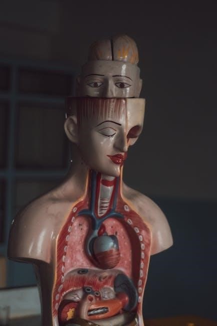
Key Systems & Their Labs
Physicians require integrated knowledge; therefore‚ medical education must provide opportunities for students to connect information across disciplines‚ enhancing comprehension.
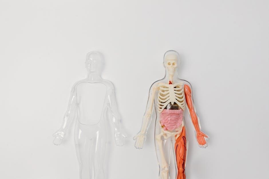
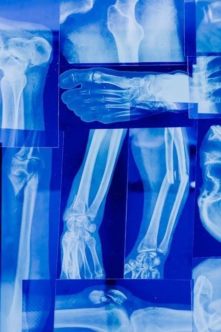
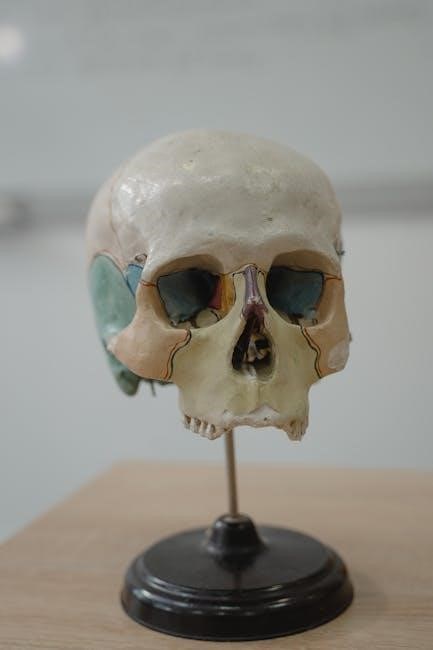
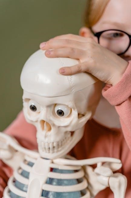
Skeletal System Lab: Bone Identification & Articulations
Understanding skeletal structures is paramount in anatomy and physiology labs‚ demanding precise bone identification skills. Students must master recognizing various bones – cranial‚ vertebral‚ appendicular – and their unique features‚ like processes‚ foramina‚ and markings.
Labs frequently involve articulating skeletal elements‚ reconstructing joints to visualize range of motion and biomechanics. This hands-on approach reinforces comprehension of articulation types: fibrous‚ cartilaginous‚ and synovial. Accurate identification of specific articulations‚ such as the shoulder or knee‚ is crucial.
Furthermore‚ labs often require classifying bones by shape – long‚ short‚ flat‚ irregular – and understanding their roles in support‚ protection‚ and movement. Successful completion necessitates diligent study and practical application of anatomical knowledge.
Muscular System Lab: Muscle Identification & Function
Mastering muscular anatomy requires identifying superficial and deep muscles‚ understanding their origins‚ insertions‚ and actions. Lab exercises often involve palpating muscles on anatomical models or cadavers‚ reinforcing spatial relationships and tactile learning.
Students must correlate muscle names with their functions – flexion‚ extension‚ abduction‚ adduction – and analyze how muscle groups work synergistically or antagonistically to produce movement. Dissections may reveal muscle fiber arrangements and fascicle structures‚ linking anatomy to physiological performance.
Labs also emphasize understanding muscle contraction mechanisms and the role of neurotransmitters. Accurate identification of specific muscles‚ like the biceps brachii or gastrocnemius‚ and their associated actions is essential for success.
Nervous System Lab: Brain Dissection & Nerve Identification
Exploring the nervous system demands meticulous brain dissection and precise nerve identification. Labs typically involve sheep brains‚ allowing students to visualize gyri‚ sulci‚ and major brain regions – cerebrum‚ cerebellum‚ brainstem – in three dimensions.
Identifying cranial nerves and tracing their pathways is crucial‚ linking structure to function. Students learn to differentiate between gray and white matter‚ understanding their roles in information processing. Dissection reveals the intricate organization of the brain‚ highlighting its complexity.
Labs also focus on spinal cord anatomy and reflex arcs‚ demonstrating neural pathways. Accurate identification of brain structures and nerves is paramount for understanding neurological processes and potential pathologies.
Cardiovascular System Lab: Heart Dissection & Blood Vessel Anatomy
Cardiovascular labs center around heart dissection‚ revealing chambers‚ valves‚ and major vessels. Sheep hearts are commonly used‚ providing a tangible model for understanding cardiac anatomy. Students identify the aorta‚ vena cava‚ pulmonary artery‚ and coronary arteries‚ tracing blood flow pathways.
Dissection emphasizes the relationship between structure and function‚ illustrating how valve placement ensures unidirectional blood flow. Examining the heart wall layers – epicardium‚ myocardium‚ endocardium – clarifies tissue composition. Labs extend to blood vessel anatomy‚ differentiating arteries‚ veins‚ and capillaries.
Understanding vessel structure – tunica intima‚ media‚ and adventitia – is vital; Accurate identification of these components reinforces comprehension of circulatory dynamics and potential cardiovascular diseases.
Respiratory System Lab: Lung Anatomy & Breathing Mechanics
Respiratory system labs focus on lung dissection and understanding breathing mechanics. Sheep lungs are frequently utilized‚ allowing students to visualize lobar structure‚ bronchi‚ and bronchioles. Identifying the trachea‚ larynx‚ and diaphragm is crucial for comprehending air passage and the mechanics of inspiration/expiration.
Labs often involve demonstrating lung volume measurements‚ such as tidal volume and vital capacity‚ using spirometers. This reinforces the physiological principles governing airflow. Observing alveolar structure under a microscope highlights gas exchange surfaces.
Understanding the role of intercostal muscles and the diaphragm in altering thoracic cavity volume is key. Relating anatomical structures to their functional roles solidifies comprehension of respiratory physiology.
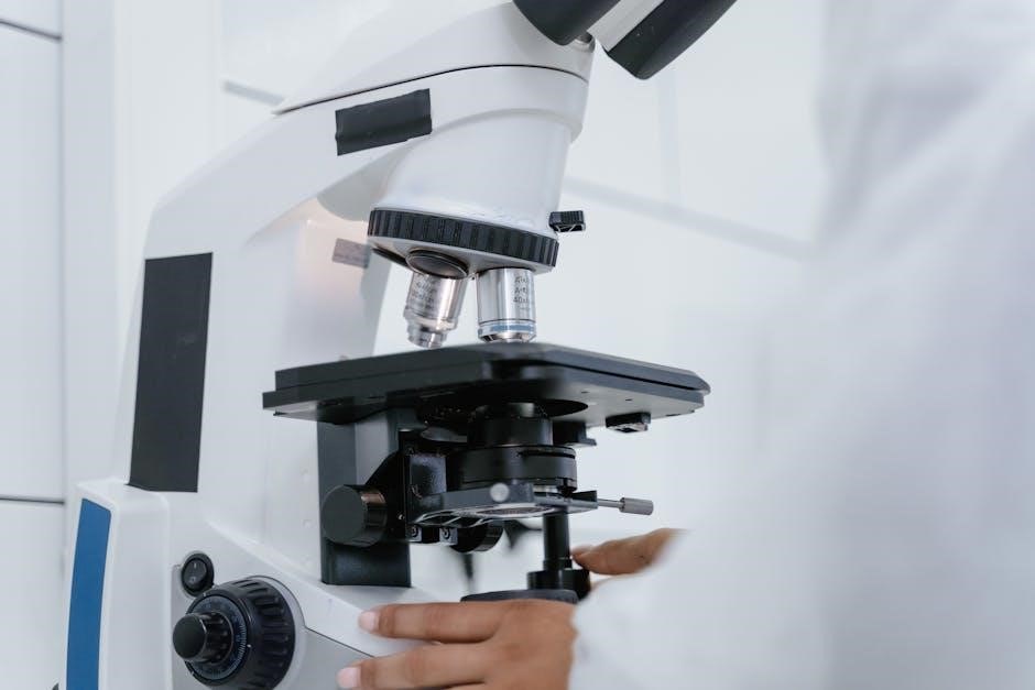
Microscopy & Histology
Microscopic examination of tissues is vital; preparing slides and identifying tissue types—epithelial‚ connective‚ muscle‚ and nervous—enhances understanding of anatomical structures.
Preparing Microscope Slides
Effective slide preparation is fundamental to histological study‚ demanding meticulous technique for clear visualization. Begin with a thin tissue section‚ carefully placed on a glass slide. Apply a fixative‚ like formalin‚ to preserve cellular structure and prevent decomposition‚ ensuring long-term integrity for observation.
Next‚ dehydration through a series of alcohol solutions removes water‚ preparing the tissue for embedding in paraffin wax‚ providing support during sectioning. A microtome then slices the wax-embedded tissue into incredibly thin sections – typically 5-10 micrometers – allowing light transmission.
These sections are mounted onto slides and stained with dyes‚ such as hematoxylin and eosin (H&E)‚ to highlight different cellular components. Hematoxylin stains nuclei blue‚ while eosin stains cytoplasm pink‚ creating contrast for detailed analysis. Finally‚ a coverslip is carefully applied‚ protecting the specimen and enhancing image clarity under the microscope.
Identifying Tissue Types Under the Microscope
Microscopic tissue identification relies on recognizing distinct structural characteristics. Epithelial tissue appears as closely packed cells‚ forming coverings and linings; observe cell shape (squamous‚ cuboidal‚ columnar) and layering (simple‚ stratified). Connective tissue‚ abundant and diverse‚ showcases varied cell types and extracellular matrices – identify fibers like collagen and elastin.
Muscle tissue exhibits contractile filaments; distinguish skeletal (striated‚ multinucleated)‚ smooth (non-striated‚ spindle-shaped)‚ and cardiac (striated‚ branched) types. Nervous tissue comprises neurons and glial cells; recognize neuron cell bodies‚ axons‚ and dendrites.
Staining techniques‚ like H&E‚ enhance visualization. Hematoxylin highlights nuclei‚ aiding in cell identification‚ while eosin stains cytoplasm‚ revealing cellular details. Careful observation of these features‚ coupled with practice‚ builds proficiency in histological analysis.
Physiological Measurements & Analysis
Accurate measurements of heart rate‚ blood pressure‚ and respiratory volumes are crucial for understanding bodily functions and assessing physiological responses effectively.
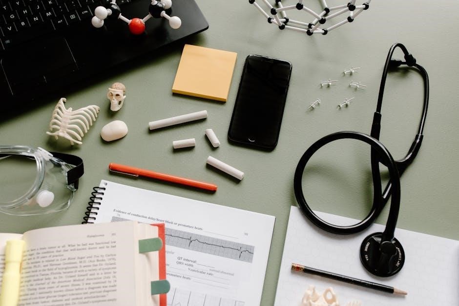
Measuring Heart Rate & Blood Pressure
Precisely determining heart rate and blood pressure are fundamental physiological assessments within anatomy and physiology labs. Students learn techniques like auscultation using a stethoscope to count heartbeats per minute‚ establishing baseline values and observing responses to exercise or stimuli.
Blood pressure measurement typically employs a sphygmomanometer‚ recording systolic and diastolic pressures – vital indicators of cardiovascular health. Understanding the factors influencing these measurements‚ such as posture‚ stress‚ and medication‚ is essential.
Analyzing variations from normal ranges allows for insights into potential health concerns. Labs often involve repeated measurements and calculations to assess individual physiological responses and reinforce understanding of circulatory system dynamics. Proper technique and accurate recording are paramount for reliable data analysis.
Analyzing Respiratory Volumes
Respiratory volume analysis is a cornerstone of physiology labs‚ enabling students to quantify lung function. Techniques often involve spirometry‚ utilizing a spirometer to measure vital capacities like tidal volume (TV)‚ inspiratory reserve volume (IRV)‚ expiratory reserve volume (ERV)‚ and residual volume (RV).
Calculating derived values‚ such as total lung capacity (TLC) and minute ventilation‚ provides a comprehensive assessment of respiratory efficiency. Students learn to correlate these volumes with factors like age‚ sex‚ and body size‚ recognizing normal ranges and identifying potential abnormalities.
Understanding lung mechanics and the impact of conditions like asthma or emphysema is reinforced through data interpretation. Accurate measurement and careful analysis are crucial for comprehending the physiological basis of breathing and gas exchange.
Utilizing Online Resources & Study Aids
Nursing Times offers over 6‚000 peer-reviewed clinical articles‚ learning units‚ and an AI-powered tool‚ enhancing anatomy & physiology understanding for students.
Reputable Online Anatomy & Physiology Resources
Accessing reliable online resources is crucial for supplementing lab manual studies. Nursing Times provides a wealth of information‚ boasting over 6‚000 peer-reviewed clinical articles‚ offering in-depth explorations of physiological processes and anatomical structures. Their exclusive learning units present complex concepts in a digestible format‚ ideal for reinforcing lab work.
Furthermore‚ the AI-powered Ask Nursing Times tool offers immediate assistance with challenging questions‚ acting as a virtual tutor. Beyond specific answers‚ these resources emphasize the integration of knowledge across disciplines – a skill vital for future healthcare professionals. Educators should encourage students to utilize such platforms to cognitively connect information learned in the lab with broader medical contexts‚ fostering a more holistic understanding of anatomy and physiology.
Effective Study Strategies for Lab Practical Exams
Mastering lab practical exams requires more than just memorization; it demands a deep understanding of anatomical relationships and physiological functions. Utilize the comprehensive resources available‚ like those offered by Nursing Times‚ to reinforce concepts beyond the lab manual. Focus on actively applying knowledge – don’t just read about muscle origins and insertions‚ physically trace them on a model.
Employ spaced repetition‚ revisiting material at increasing intervals‚ to solidify long-term retention. Create flashcards‚ focusing on both identification and function. Practice dissecting (or virtually dissecting) structures repeatedly. Finally‚ simulate exam conditions by timing yourself and working through practice scenarios. Remember‚ integrating knowledge across disciplines‚ as emphasized by medical educators‚ will significantly enhance performance.
Addressing Common Lab Manual Challenges
Navigating anatomy and physiology lab manuals often presents hurdles. Students frequently struggle with complex terminology‚ intricate diagrams‚ and the sheer volume of information. A key challenge is translating textbook knowledge into practical identification skills. Resources like Fundamentals of Anatomy & Physiology (Martini‚ Nath‚ Bartholomew) can provide a solid foundation‚ but supplemental study is crucial.
Difficulty arises when applying theoretical concepts to real-world dissections or microscopic slides. Utilizing online resources‚ such as those from Nursing Times‚ offers alternative explanations and visual aids. Peer collaboration and seeking clarification from instructors are invaluable. Remember‚ consistent review and active learning are essential to overcome these common obstacles and build confidence.